PRAME (EP461) Rabbit Monoclonal Primary Antibody
Specialties: Dermatopathology
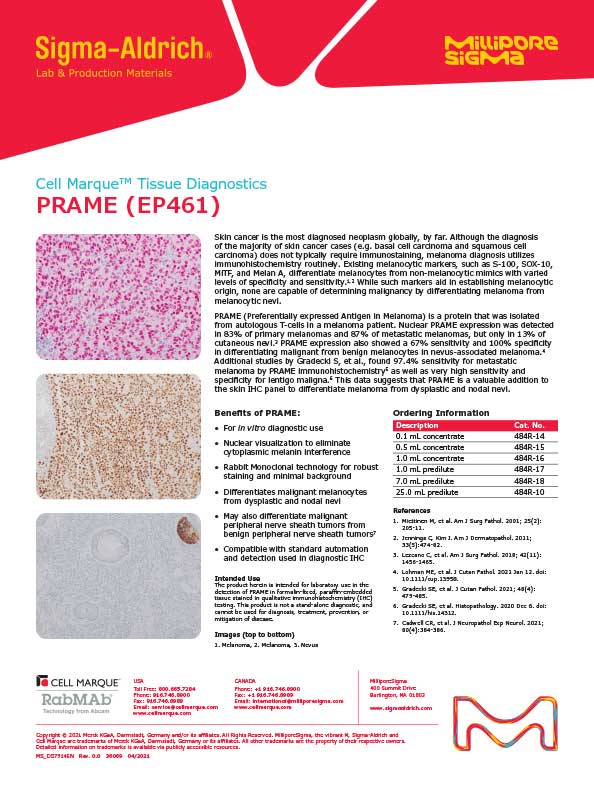
Detection of PRAME by immunohistochemistry (IHC) with PRAME (EP461) Rabbit Monoclonal Primary Antibody may be used to aid in the diagnosis of melanomas.
PRAME, or preferentially expressed antigen in melanoma, is a member of the cancer-testis antigens family and represses retinoic acid-induced cell proliferation arrest, differentiation, and apoptosis.1-3 PRAME expression is absent in most normal tissues including melanocytes and squamous epithelium, but is seen normally in testis, ovary, placenta, adrenal gland, sebaceous glands, and endometrium. It is frequently overexpressed in melanomas and overexpression has been found in other malignancies as well.2-7 PRAME immunohistochemistry has been used to differentiate melanomas from benign melanocytic proliferations such as nevi, and shows a nuclear pattern of staining.2-4
- Wadelin, Frances et al. “Leucine-rich repeat protein PRAME: expression, potential functions and clinical implications for leukaemia.†Molecular cancer vol. 9 226. 27 Aug. 2010.
- Lezcano, Cecilia et al. “PRAME Expression in Melanocytic Tumors.†The American journal of surgical pathology vol. 42,11 (2018): 1456- 1465.
- Ikeda, H et al. “Characterization of an antigen that is recognized on a melanoma showing partial HLA loss by CTL expressing an NK inhibitory receptor.†Immunity vol. 6,2 (1997): 199-208.
- Lezcano, Cecilia et al. “Immunohistochemistry for PRAME in the Distinction of Nodal Nevi From Metastatic Melanoma.†The American journal of surgical pathology vol. 44,4 (2020): 503-508.
- Zhang, Wa et al. “PRAME expression and promoter hypomethylation in epithelial ovarian cancer.†Oncotarget vol. 7,29 (2016): 45352- 45369.
- Gezgin, Gülçin et al. “PRAME as a Potential Target for Immunotherapy in Metastatic Uveal Melanoma.†JAMA ophthalmology vol. 135,6 (2017): 541-549.
- Toyama, Aimi et al. “Analyses of molecular and histopathologic features and expression of PRAME by immunohistochemistry in mucosal melanomas.†Modern pathology : an official journal of the United States and Canadian Academy of Pathology, Inc vol. 32,12 (2019): 1727-1733.
- Nadji M, Morales AR. Immunoperoxidase, part I: the techniques and its pitfalls. Lab Med 1983;14:767
- Omata M, Liew CT, Ashcavai M, Peters RL. Nonimmunologic binding of horseradish peroxidase to hepatitis B surface antigen. A possible source of error in immunohistochemistry. Am J Clin Pathol. 1980 May; 73(5):626-32. doi: 10.1093/ajcp/73.5.626. PMID: 6155065.
Specifications
- Reactivity: paraffin
- Control: Melanoma (Nuclear)
- Dilution Range: 1:25-1:50 *
Package Inserts
IFU
- IVD Rev. 2.0 (CMC48431020)
Have a different keycode?
Click Here
Learn how to obtain your SDS
Ordering Information
For in vitro diagnostic (IVD) use in USA
| 25.0 mL predilute ready-to-use | 484R-10 |
| 0.1 mL concentrate | 484R-14 |
| 0.5 mL concentrate | 484R-15 |
| 1.0 mL concentrate | 484R-16 |
| 1.0 mL predilute ready-to-use | 484R-17 |
| 7.0 mL predilute ready-to-use | 484R-18 |
For research use only (RUO) in Canada
| 25.0 mL predilute ready-to-use (RUO) | 484R-10-RUO |
| 0.1 mL concentrate (RUO) | 484R-14-RUO |
| 1.0 mL concentrate (RUO) | 484R-16-RUO |
| 1.0 mL predilute ready-to-use (RUO) | 484R-17-RUO |
| 7.0 mL predilute ready-to-use (RUO) | 484R-18-RUO |
For in vitro diagnostic (IVD) use in Europe
| 25.0 mL predilute ready-to-use | 484R-10 |
| 0.1 mL concentrate | 484R-14 |
| 0.5 mL concentrate | 484R-15 |
| 1.0 mL concentrate | 484R-16 |
| 1.0 mL predilute ready-to-use | 484R-17 |
| 7.0 mL predilute ready-to-use | 484R-18 |
For research use only (RUO) in Japan
| 25.0 mL predilute ready-to-use (RUO) | 484R-10-RUO |
| 0.1 mL concentrate (RUO) | 484R-14-RUO |
| 1.0 mL concentrate (RUO) | 484R-16-RUO |
| 1.0 mL predilute ready-to-use (RUO) | 484R-17-RUO |
| 7.0 mL predilute ready-to-use (RUO) | 484R-18-RUO |
To request information on this product in additional countries, please click the button below.