Napsin A (EP205) Rabbit Monoclonal Antibody
Specialties: Cytopathology Pulmonary Pathology
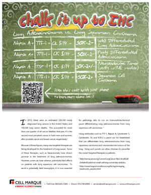
Napsin is a pepsin-like aspartic proteinase in the A1 clan of the AA clade of proteinases.1-3 There are two closely related napsins, napsin A (NAPSA) and napsin B (NAPSB).1-3 Napsin A is involved in processing propeptide pulmonary surfactant protein B (proSP-B) in the lung.4 In normal tissue, Napsin A is expressed in type II pneumocytes of the lung and proximal tubules of the kidney.1-3 Napsin A is a useful marker for lung adenocarcinoma.1-3, 5-8
Note: This product is in obsolescence. Napsin A (MRQ-60) is available as an alternative.
- Jagirdar J. Application of immunohistochemistry to the diagnosis of primary and metastatic carcinoma to the lung. Arch Pathol Lab Med. 2008; 132:384-96.
- Bishop JA, et al. Napsin A and thyroid transcription factor-1 expression in carcinomas of the lung, breast, pancreas, colon, kidney, thyroid, and malignant mesothelioma. Hum Pathol. 2010; 41:20-5.
- Rawlings ND and Salvesen GS. Handbook of Proteolytic Enzymes Volume 1. 3rd Edition. Academic Press. 2013; p.69-71.
- Brasch F, et al. Involvement of napsin A in the C- and N-terminal processing of surfactant protein B in type-II pneumocytes of the human lung. J Biol Chem. 2003; 278: 49006-14.
- Dejmek A, et al. Napsin A (TA02) is a useful alternative to thyroid transcription factor-1 (TTF-1) for the identification of pulmonary adenocarcinoma cells in pleural effusions. Diagn Cytopathol. 2007; 35:493-7.
- Inamura K, et al. Pulmonary adenocarcinomas with enteric differentiation: histologic and immunohistochemical characteristics compared with metastatic colorectal cancers and usual pulmonary adenocarcinomas. Am J Surg Pathol. 2005; 29:660-5.
- Ye J, et al. Combination of Napsin A and TTF-1 immunohistochemistry helps in differentiating primary lung adenocarcinoma from metastatic carcinoma in the lung. Appl Immunohistochem Mol Morphol. 2011; 19:313-17.
- Mukhopadhyay S, et al. Subclassification of non-small cell lung carcinomas lacking morphologic differentiation on biopsy specimens: Utility of an immunohistochemical panel containing TTF-1, napsin A, p63, and CK5/6. Am J Surg Pathol. 2011; 35:15-25.
Specifications
- Reactivity: paraffin
- Visualization: cytoplasmic
- Control: lung adenocarcinoma, kidney, renal cell carcinoma
- Dilution Range: 1:100-1:500*
Package Inserts
IFU
- IVD Rev. 2.0 (CMC35231020)
Have a different keycode?
Click Here
Learn how to obtain your SDS
Ordering Information
For in vitro diagnostic (IVD) use in USA
| 0.1 mL concentrate | 352R-14 |
| 0.5 mL concentrate | 352R-15 |
| 1 mL concentrate | 352R-16 |
| 1 mL predilute | 352R-17 |
| 7 mL predilute | 352R-18 |
For in vitro diagnostic (IVD) use in Canada
| 0.1 mL concentrate | 352R-14 |
| 0.5 mL concentrate | 352R-15 |
| 1 mL concentrate | 352R-16 |
| 1 mL predilute | 352R-17 |
| 7 mL predilute | 352R-18 |
For in vitro diagnostic (IVD) use in Europe
| 0.1 mL concentrate | 352R-14 |
| 0.5 mL concentrate | 352R-15 |
| 1 mL concentrate | 352R-16 |
| 1 mL predilute | 352R-17 |
| 7 mL predilute | 352R-18 |
For research use only (RUO) in Japan
| 0.1 mL concentrate | 352R-14-RUO |
| 0.5 mL concentrate | 352R-15-RUO |
| 1 mL concentrate | 352R-16-RUO |
| 1 mL predilute | 352R-17-RUO |
| 7 mL predilute | 352R-18-RUO |
To request information on this product in additional countries, please click the button below.